
Scientific Publications Guide, Part 1
Summary points from 2 scientific publications
This series of posts reviews results at the foundation of deCervo’s products
July 1, 2023
We provide you an easy-to-read guide of the scientific publications that underlie our simple apps
Here, we look at two articles from our Publications page
How The Brain Changes After Training
We compared hitters to non-hitters to see how their brains “cool down” from a hitting workout
We begin this scientific publications guide by going line by line on the paper’s abstract. From this translation, hopefully you will have a greater appreciation of the amazing way our brains recover from a cognitively demanding task, like hitting a baseball! (For reference, read about cooling down from a physically demanding task to see how it’s different than what we describe.) Let’s begin by translating this scientific publication into an easy-to-read guide.
The first two sentences of the abstract tell us:
“Post-task resting state dynamics can be viewed as a task-driven state where behavioral performance is improved through endogenous, non-explicit learning. Tasks that have intrinsic value for individuals are hypothesized to produce post-task resting state dynamics that promote learning.”
Translation:
Some tasks you do will activate internal changes to your brain to help you learn those tasks. If you think those tasks are valuable to you then after practicing them your brain will do things to help solidify that learning.
The next sentence of the abstract tells us:
“We measured simultaneous fMRI/EEG and DTI in Division-1 collegiate baseball players and compared to a group of controls, examining differences in both functional and structural connectivity.”
Translation:
We measured brain activity of Division-1 hitters and compared them to normal male college kids. Then we looked at how/if this brain activity may be different in how these two groups use their brains. We also examined if the actual structure of these two groups’ brains are different from each other.
The next sentences tell us:
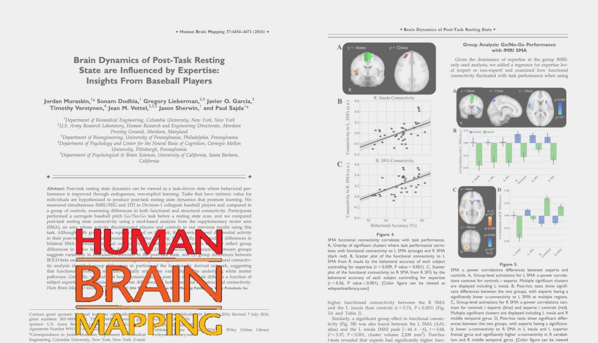
“Participants performed a surrogate baseball pitch Go/No-Go task before a resting state scan, and we compared post-task resting state connectivity using a seed-based analysis from the supplementary motor area (SMA), an area whose activity discriminated players and controls in our previous results using this task. Although both groups were equally trained on the task, the experts showed differential activity in their post-task resting state consistent with motor learning.”
Translation:
The two groups (college hitters and regular students) played the same video game. You did better at this game if you could identify different baseball pitches (like slider, curveball, fastball, etc.). Sometimes they had to “Go” (like swinging a bat) and sometimes they had to not go, or “No-Go”, to indicate they took a pitch.
They did this before what is called a “resting state scan.” “Resting state scan” means a measurement of brain activity when you are lying still and doing nothing. We then looked at the two groups’ brains when doing nothing, like this. We specifically looked at connections coming from an area called “SMA.” This is a part of the brain highly involved in how you move. Also we had seen in other papers (see here) that this part of the brain is very important for being a good hitter. We found that the college hitters had different activity in this part of the brain when just lying still.
The next sentences tell us:
“Specifically, we found (1) differences in bilateral SMA–L Insula functional connectivity between experts and controls that may reflect group differences in motor learning, (2) differences in BOLD-alpha oscillation correlations between groups suggests variability in modulatory attention in the post-task state, and (3) group differences between BOLD-beta oscillations that may indicate cognitive processing of motor inhibition.”
Translation:
We found three main differences between the groups of college hitters and regular students.
First, both sides of the “SMA” work differently in the college hitters than they do in the regular students. By “functional connectivity,” we mean the places that work in connection with the SMA. These places are connected differently in hitters vs. non-hitters.
Second, the “BOLD-alpha oscillation” is a fancy term for a type of bloodflow measurement in the brain. Specifically, we found that after a pitch, the hitters’ bloodflow relationships (or “correlations”) look different than the regular students’. This difference is likely connected to their attention on what they just did while they are resting.
Third, another type of bloodflow called “BOLD-beta oscillations” shows group differences between hitters and non-hitters. This difference is likely related to differences in how well the hitters are able to lay off bad pitches (also called “motor inhibition”).
The final sentences tell us:
“Structural connectivity analysis identified group differences in portions of the functionally derived network, suggesting that functional differences may also partially arise from variability in the underlying white matter pathways. Generally, we find that brain dynamics in the post-task resting state differ as a function of subject expertise and potentially result from differences in both functional and structural connectivity.”
Translation:
The hitters brains are wired differently than the regular students. This is especially true in the regions of the brain used to track and hit a baseball. Our general result is that the way the brains work after tracking and hitting baseballs is different for hitters and non-hitters. These differences show up in how the brain works together (“functional”) and how it is physically connected (“structural connectivity”).
Putting Together Many Ways To Measure the Brain
We found how seeing and moving are intimately tied in baseball hitters
We go to the next paper in this scientific publications guide. To understand this paper, we are again going to go line by line on the abstract. From this translation, hopefully you will have a greater appreciation of the amazing way our brains are formed. These brain connections make it possible for a seamless integration to occur between seeing a pitch and making a swing on it (watch video on this here)! Let’s begin by translating this scientific publication too into an easy-to-read guide.
The first sentence of the abstract tells us:
“In the last few decades, noninvasive neuroimaging has revealed macroscale brain dynamics that underlie perception, cognition, and action.”
Translation:
Over the last thirty years, scientists have found ways to take “pictures” of the brain doing stuff. These pictures reveal how parts of the brain interact to create what we see, think and do.
The next sentences of the abstract tell us:
“Advances in noninvasive neuroimaging target two capabilities: 1) increased spatial and temporal resolution of measured neural activity; and 2) innovative methodologies to extract brain–behavior relationships from evolving neuroimaging technology. We target the second.”
Translation:
There are two ways of taking even better brain pictures. 1) You can take more detailed “pictures” and faster “shutter speed.” 2) You can cleverly put together the brain pictures you have to link it to how people behave. In this study, we did the second.
The next sentences tell us:
“Our novel methodology integrated three neuroimaging methodologies and elucidated expertise-dependent differences in functional (fused EEG-fMRI) and structural (dMRI) brain networks for a perception–action coupling task. A set of baseball players and controls performed a Go/No-Go task designed to mimic the situation of hitting a baseball.”
Translation:
Our new approach put together three ways of taking brain “pictures.” From all three of these ways (“EEG” and “fMRI”), we could see how hitters were different than non-hitters. The differences were both in how the hitters used their brains (“functional”) and how their brains are already wired (“structural”). College-aged baseball hitters and regular college students (“controls”) played a videogame that mimicked the Go/No-Go decisions required to hit a baseball from the plate.
The next sentences tell us:
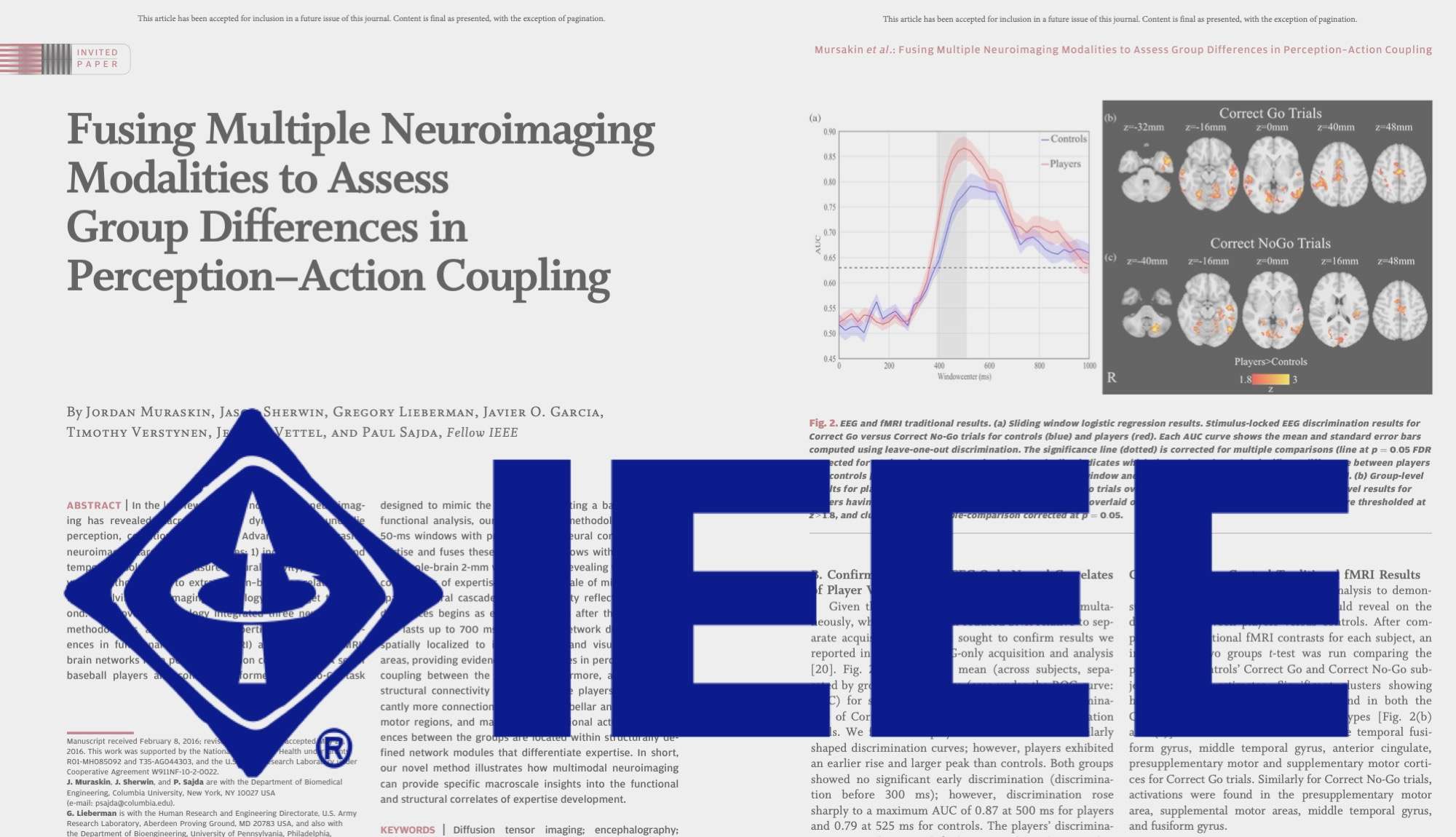
“In the functional analysis, our novel fusion methodology identifies 50-ms windows with predictive EEG neural correlates of expertise and fuses these temporal windows with fMRI activity in a whole-brain 2-mm voxel analysis, revealing time-localized correlations of expertise at a spatial scale of millimeters. “
Translation:
We analyzed how parts of the brain are working together first. To do this, we looked at the brains of hitters and non-hitters during moments in time as the ball is coming towards the plate.
We could identify from these brain “pictures” whether it was a picture of a hitter’s brain or a regular student’s. We then used that information to look at “very detailed pictures” of the brains of each group. These pictures told us exactly when and where certain parts of the brain have to be used in order to be good at hitting a baseball. We can tell what parts of the brain are relevant for hitting down to a millimeter.
The next sentence tells us:
“The spatiotemporal cascade of brain activity reflecting expertise differences begins as early as 200 ms after the pitch starts and lasts up to 700 ms afterwards.”
Translation:
There are parts and sequences of brain activity that tell us who is a good hitter and who is not. These parts and sequences reveal themselves 0.02-sec after the ball leaves the pitcher’s hand. From the pitch release until this time, you cannot tell the difference between brain activity of a college hitter and a regular student’s. From 0.02-sec, these differences last through when the ball gets to the plate.
These differences even continue after the hitter has swung or not. Based on pitch speeds we used, we saw the differences last until 0.07-sec from when the ball left the pitcher’s hand. After that, you cannot tell the difference between a hitter’s brain and a regular student’s using this method.
The next sentence tells us:
“Network differences are spatially localized to include motor and visual processing areas, providing evidence for differences in perception–action coupling between the groups.”
Translation:
The differences we found between hitters and non-hitters were specific to parts of the brain for moving and seeing.
The next sentence tells us:
“Furthermore, an analysis of structural connectivity reveals that the players have significantly more connections between cerebellar and left frontal/ motor regions, and many of the functional activation differences between the groups are located within structurally defined network modules that differentiate expertise.”
Translation:
When we looked at how the two groups’ brains were wired together, the hitters had more “wiring” between parts of the brain related to movement. When we looked at how the two groups used their brains, we found that hitter’s brains are like other people’s who are skilled at a certain task (e.g., driving, or flying).
The final sentence tells us:
“In short, our novel method illustrates how multimodal neuroimaging can provide specific macroscale insights into the functional and structural correlates of expertise development.”
Translation:
To summarize, our new way of putting together brain “pictures” shows how wiring and use are different in brains of skilled people.